Electrocardiogram

Also, known as an EKG or ECG.
Records the electrical activity from the heart.
Einthoven's Triangle

3 electrodes: positive, negative, and ground
Lead: the imaginary line created by the electrodes
5 Lead System
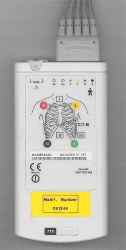
Electrode Placement
RA (white): below R clavicle, 2nd ICS, R midclavicular line.
RL (green): R Lower rib cage, 8th ICS, R midclavicular line.
LA (black): below L clavicle, 2nd ICS, L midclavicular line.
LL (red): L lower rib cage, 8th ICS, L midclavicular line.
V1 (brown): 4th ICS, R sternal border
Remember: White on Right, White skies over green pastures and Smoke (black) over fire (red).

EKG PAPER

Horizontal Axis: time and rate measured
Vertical Axis: Amplitude/ Voltage Measured
Deflections

Positive Deflection: EKG waveform that moves above the isoelectric line. (Moves Up toward the top of the paper).
Negative Deflection: EKG waveform that moves below the isoelectric line. (Moves down toward the bottom of the paper).
Biphasic Deflection: EKG waveform that moves both below and above the isoelectric line.
Negative Deflection: EKG waveform that moves below the isoelectric line. (Moves down toward the bottom of the paper).
Biphasic Deflection: EKG waveform that moves both below and above the isoelectric line.
