Waves, Complexes, and Intervals

P Wave

P Wave: 1st deflection. Electrical stimulation of the SA node, resulting in atrial contraction.
Normal appearance: smooth, round, and upright.
Normal appearance: smooth, round, and upright.
PR Interval
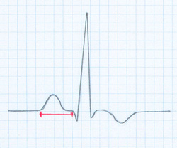
PR Interval (PRI): measurement between the beginning of the P wave to the beginning of the QRS complex.
Normal PRI: 0.12 to 0.20 second
QRS Complex
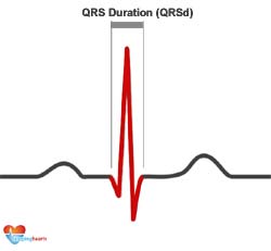
QRS Complex: Consist of 3 wave deflections immediately following the P wave. Represents electrical impulse conducted from the bundle of His to the Purkinje fibers.
Q wave:negative deflection following P wave and immediately preceding the R wave.
R wave: Positive wave form following the Q wave.
S wave: Negative deflection immediately following the Q wave.
Normal QRS: < 0.10 second
Q wave:negative deflection following P wave and immediately preceding the R wave.
R wave: Positive wave form following the Q wave.
S wave: Negative deflection immediately following the Q wave.
Normal QRS: < 0.10 second
Q Wave

Q Wave: negative deflection following P wave and immediately preceding the R wave. Deep, wide Q waves may indicate myocardial infarction.
ST segment

ST Segment: Represents the end of ventricular depolarization. Normal deflection: baseline or isoelectric line.
Elevated/ depressed= myocardial injury/ ischemia
Elevated/ depressed= myocardial injury/ ischemia
T Wave

T Wave: Follows ST segment. Ventricular repolarization or ventricular recovery phase (resting state).
